Examples of Enhancing Treatment & Care with HBOT: Golfer
Case: Golfer the lab lost his tail wag – acute disk extrusion at sacrococcygeal junction
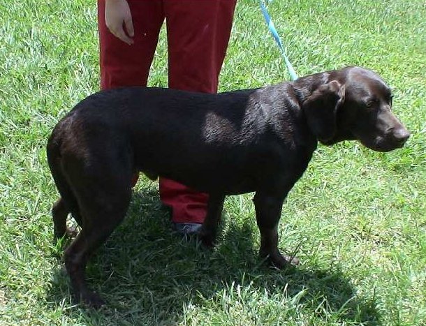
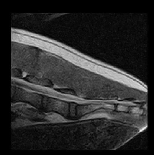
Above left: Golfer. Above right: MRI
A 2 year old M/N Lab was examined for acute onset of inability to use or wag his tail. The owner reported that the day before presentation the dog had spent hours playing vigorously with several other canine friends. The following morning the owner awoke to find the dog unable to use his tail. The tail was held limp between the rear legs, and the Lab was periodically biting at it. The primary care veterinarian referred the case immediately after exam and prior to any treatment. The dog was able to urinate and defecate normally with complete continence.
Neurologic exam revealed normal mentation, cranial nerve function, gait, forelimb and rear limb proprioceptive positioning and reflexes. Anal reflex and tone were normal. The tail was limp, with only slight movement at the base, and poor tone. Sensation was intact to all areas of the tail.
Color Doppler Ultrasound exam revealed normal blood flow to the tail vasculature. MR Imaging revealed a dorsal attenuation of the coccygeal canal at S3-Cy1. He was diagnosed with an acute disk extrusion at the sacrococcygeal junction causing tail paralysis.
Treatment offered included medical with hyperbaric oxygen therapy. Depending on response, surgery would be offered within 3-5 days if no improvement noted.
The patient was started on Tramadol 50mg BID and Gabapentin 100mg BID. Hyperbaric Oxygen Therapy at 1.5 ATA BID was initiated.
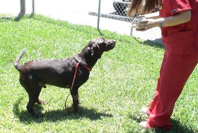
Outcome: Within 36 hours the patient was lifting and moving his tail. By day 3, the “Happy Golfer” and his wiggly tail had returned.
HBOT Schedule
Day 1: 1 Treatment at 1.5 ATA
Day 2: 2 Treatments at 1.5 ATA
Day 3: 1 Treatment at 1.5 ATA
© RKLyman, LLC. All Rights Reserved.